Plasminogen deficiency Type 1 (plgd-1)
SYMPTOMS & SIGNS

Graphic representation of plasminogen deficiency symptoms presenting as woody eye growths

8 out of 10
Individuals with
ligneous conjunctivitis

have woody lesions in other parts of
the body1*

Lesions can impact mucous
membranes throughout the body1,2

Illustration showing how plasminogen deficiency symptoms may present as growths on the mucous membranes of the brain, ears, eyes, mouth, respiratory tract, renal system, gastrointestinal system, female genital tract, and skin

Prevalence data continue to evolve and may change as additional PLGD-1 cases are identified and more research becomes available.

Patient pool of 102 individuals with a mean age at onset of 5 years and 5 months and a consanguinity rate of 40%1

Patient pool of 74 individuals with a median age at onset of 9.5 months2

Manifestations of
PLGD-1

Oral involvement is a common manifestation of PLGD-1 with 32% of patients impacted by these thick, nodular masses on the gums with a distinctly hard, "woody" texture. This condition is known as periodontal disease, ligneous periodontitis, or ligneous gingivitis.1,9,10

With ligneous gingivitis, the gingiva often looks swollen, thickened, firm, and pale, with a characteristic yellowish-white color. Chronic irritation from fibrin deposits and ongoing inflammation can make the gums bleed easily. Over time, persistent fibrin accumulation and inflammation may destroy periodontal tissues, resulting in tooth loss and resorption of the surrounding bone.9,10

Review the lesion image gallery for more examples of ligneous gingivitis.

Ligneous gingivitis caused by congenital plasminogen deficiency type 1, manifesting as gum lesions

Ligneous cervicitis is the most commonly reported pathology of PLGD-1 in the female genital tract with dysmenorrhea as the most common presenting symptom.7 Symptoms of female genital tract involvement include vaginal discharge, abnormal bleeding, very painful menstruation, pelvic pain, and discomfort during intercourse. If untreated, these lesions may lead to infertility.3,8

While the cervix is most commonly impacted, there have been reports of involvement of the vagina, fallopian tubes, ovaries, and endometrium.7

When PLGD-1 impacts the skin, it manifests as small, translucent, yellow-brown bumps or papules on areas exposed to the sun. These lesions heal slowly, often appear in children, and are commonly known as juvenile colloid milium.7

In people with PLGD-1, wound healing capabilities are also significantly diminished because plasminogen plays a key role in tissue remodeling and wound repair. This means patients with PLGD-1 may experience impaired healing responses, which are often most evident on mucous membranes such as the conjunctivae, but also on the skin.2

In a few rare cases, children who have PLGD-1 are born with congenital obstructive hydrocephalus, a buildup of fluid in the skull.6

This occurs when lesions obstruct the flow of cerebrospinal fluid that surrounds and protects the brain and spinal cord. When cerebrospinal fluid accumulates, the extra fluid puts pressure on surrounding tissue. Surgery is often needed to relieve the obstruction.2,6

A deformity of the cerebellum, known as Dandy-Walker syndrome, can also occur in people with PLGD-1; it is more common in children but can affect adults as well.6

Lesions in the respiratory tract are among the most serious complications of PLGD-1. They may initially present as chronic cough, dysphonia, aphonia, stridor, wheezing, dyspnea, shortness of breath, and hypoxemia. Lesions can also affect the vocal cords, nasal and sinus linings, and other areas of the respiratory tract, leading to voice loss and recurrent respiratory infections.3

When they develop in the windpipe (trachea or bronchi), they can cause severe problems and may compromise and/or obstruct airways, resulting in chronic lung disease, bronchiectasis, and, in severe cases, respiratory arrest.3

Hear Wren's story

Ligneous lesions vary widely in appearance4

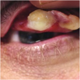
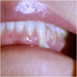
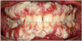
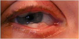
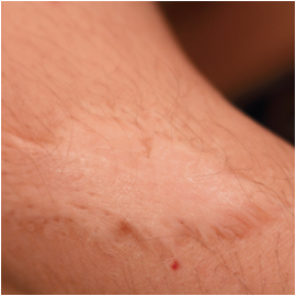
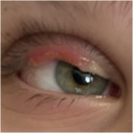
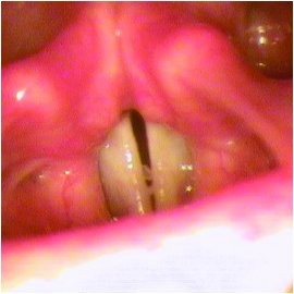
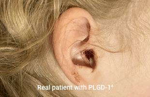
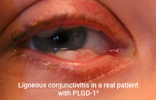
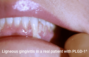
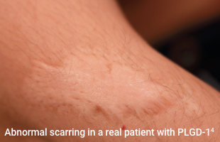
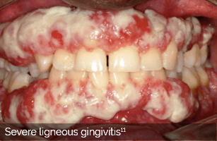
Lesions due to PLGD-1 may present in a variety of ways, from watery, stringy pseudomembranes to thick wood-like lesions. Not only does symptom severity fluctuate over time, but the overall severity of the disorder varies greatly from patient to patient depending on the location and duration of lesions. Even members of the same family impacted by PLGD-1 can present with drastically different manifestations.6

Lesions can form spontaneously, but they are typically triggered by local infection, irritation, injury, or surgical intervention.6

Explore Image Gallery >

*Patient pool primarily originates from Turkey and various Arabian countries (61%). When compared to previously published cases, this group of patients presented at a younger mean age (1 year and 10 months old) and shows a high rate of consanguinity (61%).

Diagnosing PLGD-1

Is done with a simple blood test. Undiagnosed patients may be at risk of severe complications from untreated lesions.6,7

References: 1. Klammt J, Kobelt L, Aktas D, et al. Identification of three novel plasminogen (PLG) gene mutations in a series of 23 patients with low PLG activity. Thromb Haemost. 2011;105(3):454-460. 2. Schuster V, Hügle B, Tefs K. Plasminogen deficiency. J Thromb Haemost. 2007;5(12):2315-2322. 3. Shapiro AD, Nakar C. How I treat type 1 plasminogen deficiency. Blood. 2025;145(25):2954-2965. 4. Data on file. Kedrion Biopharma Inc. 5. Balram B, Thiesen A, Kroeker KI. Inflammatory bowel disease: a gastrointestinal presentation of congenital plasminogen deficiency. ACG Case Rep J. 2021;8(5):e00613. 6. Congenital type 1 plasminogen deficiency. NORD. Updated February 6, 2025. Accessed July 16, 2025. https://rarediseases.org/rare-diseases/congenital-plasminogen-deficiency 7. Mehta R, Shapiro AD. Plasminogen deficiency. Haemophilia. 2008;14(6):1261-1268. 8. Shapiro AD, Menegatti M, Palla R, et al. An international registry of patients with plasminogen deficiency (HISTORY). Haematologica. 2020;105(3):554-561. 9. Alberto P, Cesar AV, Dayana M, et al. Ligneous gingivitis: a systematic review. J Dent Oral Sci. 2022;4(3):1-14. 10. Schuster V, Seregard S. Ligneous conjunctivitis. Surv Ophthalmol. 2003;48(4):369-388. 11. Sadasivan A, Ramesh R, Mathew DG. Ligneous periodontitis in a patient with type 1 plasminogen deficiency: a case report and review of the literature. Case Rep Dent. 2020;2020:5680535.